Biological microscopes are a cornerstone of scientific research, offering unparalleled insights into the intricate world of cells, tissues, and microorganisms. These sophisticated optical instruments are designed with precision to cater to the specific needs of biological observation, making them indispensable in various fields such as medical research, microbiology, and education.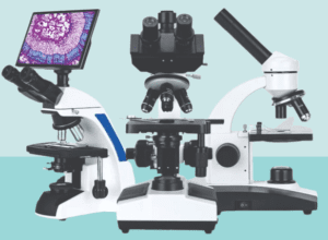
Key Components and Their Functions:
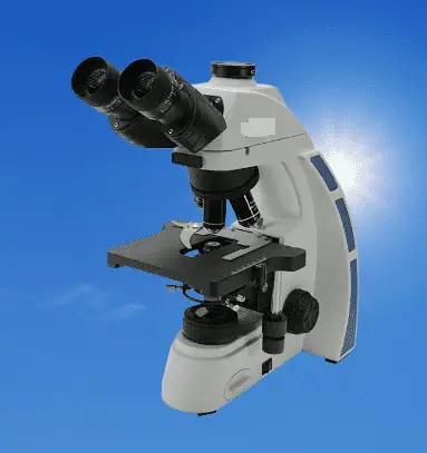
A biological microscope is composed of several critical parts, each contributing to its overall functionality:
- Eyepieces: These are used to observe the magnified image of the specimen.
- Objective Lenses: These lenses provide the primary magnification, ranging from 10x to 1,000x or more, allowing for detailed examination of biological samples.
- Stage: This is where the sample is placed. It often includes clips or other fixtures to secure the specimen.
- Condenser: This component focuses light onto the specimen, enhancing the clarity of the image.
- Diaphragm: It controls the amount of light reaching the specimen, optimizing visibility and contrast.
- Light Source: Essential for illuminating the sample, making it visible under the microscope.
Modern advancements have significantly enhanced these basic components. For instance, stages now often feature mechanical or automated designs that can be operated via a mouse, and digital cameras have replaced traditional eyepieces, allowing images to be displayed on screens for group viewing and further analysis.
How Biological Microscopes Work:
The operation of a biological microscope involves a meticulous process of light manipulation. Light from the illumination source is directed onto the specimen through the condenser. This light then passes through the objective lens, where it is magnified. Finally, the magnified light travels through the eyepieces, further enlarging the image for the viewer.
The selection of a biological microscope depends on several factors, including magnification, resolution, illumination, and operability:
- Magnification: Different samples require varying levels of magnification. For example, observing bacteria typically necessitates high magnification (100x – 150x).
- Resolution: Higher magnification usually correlates with better resolution, allowing finer details to be observed.
- Illumination/Observation: Uniform illumination and accurate color representation are crucial. Advanced models may offer phase contrast and fluorescence imaging for enhanced visualization of biological processes.
- Operability: Ease of use is important. Basic models require manual adjustments, while advanced microscopes feature automated settings controlled via mouse or touchscreen, often optimizing themselves for the sample.

Working Principle of the Biological Microscope:
A biological microscope operates on the principles of optical magnification and illumination to enable detailed observation of microscopic specimens. The fundamental working process involves several steps, each contributing to the creation of a clear and magnified image of the biological sample.
Steps in the Working Principle
- Illumination
- The microscope has a built-in light source, typically an LED or halogen lamp, that provides the necessary illumination.
- The light is directed upwards through the condenser, which focuses and concentrates the light onto the specimen.
- Specimen Placement
- The biological specimen is placed on a glass slide and positioned on the stage.
- The stage often includes clips or a mechanical holder to secure the slide in place.
- Condenser and Diaphragm Adjustment
- The condenser lens system adjusts the light to focus it on the specimen.
- The diaphragm, usually an iris or disc type, controls the amount of light and improves contrast by reducing or increasing the light intensity.
- Magnification through Objective Lenses
- Light passing through the specimen enters the objective lens.
- Objective lenses are available in various magnifications (e.g., 4x, 10x, 40x, 100x).
- The objective lens magnifies the image of the specimen. The degree of magnification depends on the lens in use.
- Formation of Real Image
- The objective lens forms a real, inverted image of the specimen within the microscope’s body tube.
- This real image is further magnified by the eyepiece lens or projected onto a digital camera sensor in advanced models.
- Magnification through Eyepieces
- The eyepieces, or ocular lenses, provide additional magnification (typically 10x or 15x).
- The user views the magnified image through the eyepieces, or it can be displayed on a screen if a digital camera is used.
- Focus Adjustment
- Coarse and fine focus knobs are used to bring the specimen into sharp focus.
- Coarse focus allows for general adjustments, while fine focus provides precise control for high magnification lenses.
- Image Observation and Analysis
- The final magnified image is observed through the eyepieces or displayed on a monitor for digital models.
- Imaging software can be used to capture, analyze, and process the image further in digital microscopes.
Technical Specifications of a Biological Microscope:
| Component | Specification |
| Eyepieces | Widefield eyepieces, typically 10x or 15x magnification |
| Objective Lenses | Multiple objective lenses (e.g., 4x, 10x, 40x, 100x) |
| Magnification Range | Total magnification ranging from 40x to 1,000x or higher (depending on eyepiece and objective combination) |
| Stage | Mechanical stage with X-Y movement; automated or manual |
| Condenser | Abbe condenser with adjustable aperture diaphragm; N.A. 1.25 or higher |
| Diaphragm | Iris diaphragm or disc diaphragm |
| Light Source | LED or halogen illumination; adjustable brightness |
| Focusing Mechanism | Coarse and fine focus adjustment knobs |
| Resolution | Dependent on objective lens quality; typically around 200 nanometers with high-quality lenses |
| Optical System | Infinity-corrected optical system for advanced models |
| Illumination Type | Brightfield, phase contrast, and fluorescence imaging options available |
| Digital Integration | Optional digital camera with high resolution (e.g., 5 MP or higher) |
| Display | LCD display or monitor for digital models |
| Software | Imaging software for capturing, analyzing, and processing images |
| Working Distance | Short working distance for high magnification lenses; varies by lens |
| Numerical Aperture | High numerical aperture (e.g., 0.65 to 1.25) for better resolution and brightness |
| Compatibility | Compatible with various accessories like fluorescence kits, phase contrast kits, and polarization kits |
| Build | Robust and stable construction to minimize vibrations |
Applications and uses of biological microscopes:
| Field | Applications | Uses |
| Medical Research | Study of cells and tissues to understand diseases and their effects | Developing new drugs, treatments, and therapies; observing drug performance under various conditions |
| Fluorescence imaging for clinical drug trials | Visualizing changes in specimens in different environments | |
| Microbiology | Study of bacteria and other microorganisms | Understanding structure and function; exploring interactions with environments |
| Education | Teaching biology and life sciences in schools | Helping students learn about cell structures and biological processes |
| Use in universities and research institutions | Training future scientists and conducting research |
Frequently Asked Questions:
What is a biological microscope?
Answer: A biological microscope is a type of optical microscope designed primarily for observing cells, tissues, and other biological specimens. It utilizes multiple objective lenses to provide varying magnifications, typically ranging from 10x to 1,000x or more. These microscopes are equipped with features like a condenser, diaphragm, and light source to create optimal viewing conditions for flat samples such as slides and petri dishes. Advanced models may include digital cameras and automated stages for enhanced functionality.
What are the 4 types of microscopes?
Answer: The four main types of microscopes are:
- Compound Microscope: Uses multiple lenses to achieve high magnification for observing small specimens like cells and bacteria.
- Stereoscopic Microscope: Provides a 3D view of larger, opaque specimens with lower magnification.
- Confocal Microscope: Uses laser light to scan specimens and produce high-resolution images with depth selectivity.
- Electron Microscope: Uses beams of electrons for extremely high magnification and resolution, suitable for observing ultrastructural details.
What is the principle of the biological microscope?
Answer: The principle of a biological microscope is based on optical magnification and illumination. It involves focusing light from an illumination source onto the specimen using a condenser. The light passes through the specimen and enters the objective lens, where it is magnified. This magnified image is further enlarged by the eyepiece lens or projected onto a digital camera sensor. Coarse and fine focus adjustments are used to obtain a sharp and clear image of the specimen.
What are the 7 types of microscope?
Answer: The seven types of microscopes are:
- Compound Microscope
- Stereoscopic Microscope
- Confocal Microscope
- Electron Microscope
- Scanning Electron Microscope (SEM)
- Transmission Electron Microscope (TEM)
- Fluorescence Microscope
What are the 3 types of microscopy?
Answer: The three main types of microscopy are:
- Optical Microscopy: Uses visible light and lenses to magnify specimens.
- Electron Microscopy: Uses electron beams for higher magnification and resolution.
- Scanning Probe Microscopy: Uses a physical probe to scan the specimen surface and create high-resolution images.
What is the biological importance of microscope?
Answer: Microscopes are biologically important because they allow scientists to observe and study cells, microorganisms, tissues, and other small biological structures that are not visible to the naked eye. This capability is crucial for understanding cellular functions, diagnosing diseases, conducting medical research, and advancing knowledge in fields like microbiology and biotechnology.
Who is the father of the microscope?
Answer: Antonie van Leeuwenhoek is often considered the father of the microscope due to his pioneering work in developing early microscopes and making significant biological discoveries with them.
What are the 10 uses of a microscope?
Answer:
- Medical Diagnosis: Identifying diseases and conditions through the examination of tissue samples and cells.
- Biological Research: Studying cell structures, functions, and interactions.
- Microbiology: Observing bacteria, viruses, and other microorganisms.
- Pathology: Examining tissue samples for abnormalities.
- Forensic Science: Analyzing evidence at a microscopic level.
- Education: Teaching biology and life sciences in schools and universities.
- Botany: Studying plant cells and tissues.
- Pharmacology: Investigating the effects of drugs on cells.
- Environmental Science: Examining samples from water, soil, and air.
- Material Science: Analyzing the microstructure of materials and surfaces.
What is the biological application of microscopy?
Answer: Biological applications of microscopy include studying cell morphology, identifying pathogens, observing cellular processes, analyzing genetic material, and conducting research on biological tissues and microorganisms. Microscopy enables detailed examination and analysis that are essential for understanding biological systems and developing medical treatments.
Who created the first microscope?
Answer: The first microscope was created by Hans Lippershey, Zacharias Janssen, and Hans Janssen, Dutch spectacle makers, in the late 16th century.
What is the function of a microscope?
Answer: The function of a microscope is to magnify small objects or specimens, making them visible and allowing for detailed observation and analysis. This is achieved by using lenses to enlarge the image of the specimen and illuminate it for clear viewing.
What is the principle of SEM?
Answer: The principle of Scanning Electron Microscopy (SEM) involves scanning a focused beam of electrons across a specimen’s surface. These electrons interact with the specimen, producing signals that are collected and converted into an image. SEM provides high-resolution, three-dimensional images of the specimen’s surface topography.

What is microscopy in biology?
Answer: Microscopy in biology is the use of microscopes to observe and study biological specimens, such as cells, tissues, and microorganisms. This practice enables scientists to examine the detailed structures and functions of biological entities, aiding in research, diagnosis, and education.
How to calculate magnification?
Answer: Magnification is calculated by multiplying the magnification power of the objective lens by the magnification power of the eyepiece lens. For example, if an objective lens has a magnification of 40x and the eyepiece lens has a magnification of 10x, the total magnification is 40x * 10x = 400x.
What is the principle of the microscope?
Answer: The principle of the microscope involves the use of lenses to magnify and resolve small objects. Light (or electrons in the case of electron microscopes) passes through or interacts with the specimen, and lenses magnify the image. The magnified image is then viewed through eyepieces or displayed on a screen for observation.
What are the 12 parts of a microscope?
- Eyepiece (Ocular Lens)
- Objective Lenses
- Stage
- Condenser
- Diaphragm
- Light Source
- Arm
- Base
- Coarse Focus Knob
- Fine Focus Knob
- Nosepiece (Turret)
- Stage Clips
Which lens is used in a microscope?
Answer: Microscopes use convex lenses for both the objective and eyepiece lenses. These lenses converge light rays to form a magnified image of the specimen.
Who is the father of microscopy?
Answer: Antonie van Leeuwenhoek is considered the father of microscopy due to his development of high-quality lenses and his significant discoveries using early microscopes.
What is microscope importance?
Answer: Microscopes are important because they allow scientists to observe and study objects that are too small to be seen with the naked eye. This capability is crucial for advancing knowledge in biology, medicine, materials science, and many other fields. Microscopes enable detailed examination of cells, tissues, microorganisms, and materials, leading to discoveries and innovations.
What are the advantages of using a microscope?
- High Magnification: Allows detailed observation of small specimens.
- Resolution: Provides clear images of tiny structures.
- Versatility: Applicable in various scientific fields.
- Diagnostic Tool: Essential for medical diagnosis.
- Research: Facilitates scientific discoveries.
- Education: Enhances learning in biology and related sciences.
- Non-Destructive: Allows observation without damaging the specimen.
- Variety: Different types of microscopes for specialized applications.
- Automation: Advanced models offer automated features.
- Imaging: Digital microscopes can capture and analyze images.
What are the applications of microscope?
- Medical Diagnosis
- Biological Research
- Microbiology
- Forensic Science
- Education
- Botany
- Pharmacology
- Environmental Science
- Material Science
- Pathology
Who discovered the microscope?
Answer: Hans Lippershey, Zacharias Janssen, and Hans Janssen, Dutch spectacle makers, are credited with the invention of the first microscope in the late 16th century.
How do microscopes work?
Answer: Microscopes work by using lenses to magnify small objects. Light or electrons are directed at the specimen, and lenses focus the light or electron beams to create a magnified image. This image is viewed through eyepieces or captured by cameras for further analysis.
What are types of microscopes?
- Compound Microscope
- Stereoscopic Microscope
- Confocal Microscope
- Electron Microscope