The Gram staining is a fundamental and widely used laboratory technique in microbiology, employed to differentiate and categorize bacteria based on their cell wall characteristics. This staining method enables microbiologists to differentiate and categorize bacteria into two distinct groups: Gram-positive and Gram-negative. Here, we delve into the fundamental techniques of the Gram stain and its significance in microbiological research and diagnostics.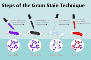
Steps of the Gram Stain technique :
| Step Number | Gram Stain Technique Step |
|---|---|
| 1 | Preparation of the Bacterial Smear |
| 2 | Fixation |
| 3 | Application of Crystal Violet |
| 4 | Iodine Treatment (Mordant) |
| 5 | Decolorization |
| 6 | Counterstaining with Safranin |
| 7 | Microscopic Examination |
| 8 | Interpretation and Analysis |
Gram staining procedure
- OBJECTIVE
- To lay down the procedure for gram staining of the given culture to identify and differentiate between gram-positive and gram-negative microorganisms.
- SCOPE
- This procedure shall be applicable for the gram staining of microorganisms in the microbiology laboratory.
- RESPONSIBILITY
- Microbiology Personnel shall be responsible for carrying out the established procedure and maintaining its records.
- Officer Microbiology or above who is trained and well versed with the procedure shall be responsible for ensuring that the procedure is carried out correctly and checking of documents for completeness of the content.
- Head – Microbiology/ Designee shall be responsible for the compliance of the SOP.
- ACCOUNTABILITY
- Head – Microbiology/ Designee shall be accountable for the proper implementation of the SOP.
- DEFINITIONS
- Gram staining: Gram staining is an empirical method of differentiating bacterial species into two large groups (Gram-positive and Gram-negative) based on the chemical and physical properties of their cell walls.
Gram Staining Procedure:
- Precautions:
- Wear a face mask and gloves before performing the test.
- Perform the preparation of the smear under the bio-safety cabinet in micro lab-2.
- Staining shall be performed in Media disposal room and observation shall be taken under microscope.
- This process is for in vitro diagnostic use only and shall be performed by adequately trained and qualified laboratory personnel.
- Decontaminate the material by autoclaving after the completion of testing.
- Materials required:
- Sterile inoculating loopGram staining kit (consisting of gram’s crystal violet, gram’s iodine, gram’s decolorizer and safranin) Sterile purified water Glass slideImmersion oil Burner Microscope.
- Note: For a thin smear, 10-20 seconds is long enough; after the proper time interval, the gram’s decolorizer drippings from the slide are no longer colored.
- Wash the slide with purified water and counter-stain it with 0.5 % ‘safranin’ for about one minute.
- Wash the slide with purified water and air dry it.
- As the slide gets completely dried put a drop of immersion oil on the smear and observe it under oil immersion objective (100X).
- Result: Gram-positive cells should stain and appear purple, while gram-negative cells should stain and appear pink colored.
- Maintain the record of gram staining as per Annexure-I.
- ABBREVIATIONS
- SOP : Standard Operating Procedure
- MB : Microbiology
- ANNEXURES
- Annexure-I : Gram Staining report
- Annexure-II : Label for Gram Staining kit receipt
GRAM STAINING REPORT
| Type of Sample | Batch No. / A.R. No. | ||
| Date | Microscope ID | ||
| Area | Batch No. / A.R. No. |
| Colony Characters on Agar plate (Sub-culture if the growth medium is broth) | ||
| Colony Characteristics | Observation | |
| Colour | ||
| Shape | Punctiform Circular Rhizoid Irregular Filamentous | |
| Pigmentation | Pigmented/ Non-pigmented | |
| Opacity | Opaque/ Transparent | |
| Elevation | ||
| Surface | Smooth/ Rough/ Wrinkled/ Dry/ Sticky | |
| Margin | Color | |
| Microscopic observation | ||
| Test | Observation | |
| Cell shape | Cocci/ Bacilli/ Coco bacilli | |
| Gram Staining | Gram Positive/ Gram Negative | |
LABEL FOR GRAM STAINING KIT RECEIPT
Steps of Gram Staining:
The Gram staining technique, a vital method in microbiology, consists of a series of sequential steps that allow for the differentiation of bacteria into Gram-positive and Gram-negative groups. Below are the key steps involved in the Gram staining technique:
- Preparation of the Bacterial Smear:
- A small sample of the bacterial culture or clinical specimen is collected and carefully spread in a thin, even layer on a clean glass slide. The smear ensures proper adhesion of the bacteria for subsequent staining.
- Fixation:
- The bacterial smear is subjected to fixation, often through heat. Passing the slide over a flame gently fixes the bacterial cells in place and prevents distortion during the staining process.
- Application of Crystal Violet:
- Crystal violet, the primary stain, is applied to the bacterial smear. The purple-colored dye permeates the cell walls of all bacterial species, regardless of their characteristics.
- Iodine Treatment (Mordant):
- Following the application of crystal violet, Gram’s iodine solution is added as a mordant. Iodine forms an insoluble complex with crystal violet within the bacterial cells, enhancing stain retention.
- Decolorization:
- The bacterial smear is then gently washed with a decolorizing agent, typically alcohol or acetone. This critical step differentiates Gram-positive and Gram-negative bacteria based on their cell wall structures.
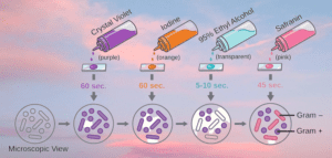
- Counterstaining with Safranin:
- After decolorization, the bacterial smear is treated with safranin, the counterstain. Gram-negative bacteria, now colorless after decolorization, readily absorb the pinkish-red hue of safranin.
- Microscopic Examination:
- The stained bacterial smear is observed under a microscope. Gram-positive bacteria retain the initial purple stain, while Gram-negative bacteria appear pinkish-red due to the uptake of safranin.
- Interpretation and Analysis:
- The microscopic examination of the stained cells allows for the differentiation and identification of bacteria as either Gram-positive or Gram-negative. This information is crucial for diagnosing infections and guiding appropriate antimicrobial treatment.
By following these step-by-step procedures, microbiologists can harness the power of the Gram staining technique to gain valuable insights into bacterial morphology and classification, contributing to both clinical diagnostics and scientific research in the field of microbiology.
Frequently Asked Questions:
What is the Gram stain technique, and why is it important in microbiology?
Answer: The Gram staining technique is a laboratory procedure used to differentiate and categorize bacteria into two groups: Gram-positive and Gram-negative. It is essential in microbiology because it provides valuable information about the cell wall characteristics of bacteria, aiding in their identification and classification.
Who developed the Gram stain technique, and when was it introduced?
Answer: The Gram staining technique was developed by Danish bacteriologist Hans Christian Gram in 1884. He introduced this staining method as a way to differentiate bacteria based on their staining reactions.
What are the primary and counterstains used in the Gram stain procedure?
Answer: The primary stain used in the Gram staining procedure is crystal violet, and the counterstain is safranin. Crystal violet stains all bacterial cells, and safranin is used to stain Gram-negative bacteria.
Describe the key steps involved in the Gram stain technique.
Answer: The Gram stain technique involves the following steps:
- Preparation of the bacterial smear
- Fixation of the smear
- Application of crystal violet
- Iodine treatment (mordant)
- Decolorization with alcohol or acetone
- Counterstaining with safranin
- Microscopic examination of stained cells
- Interpretation of the results.
How does the Gram stain differentiate Gram-positive and Gram-negative bacteria?
Answer: The key differentiating factor is the cell wall structure of bacteria. Gram-positive bacteria have a thick peptidoglycan layer that retains the crystal violet-iodine complex, making them appear purple. Gram-negative bacteria have a thinner peptidoglycan layer and lose the crystal violet-iodine complex during decolorization, causing them to appear pinkish-red after counterstaining.
What is the role of Gram’s iodine in the Gram stain procedure?
Answer: Gram’s iodine acts as a mordant, forming an insoluble complex with crystal violet within the bacterial cells. This enhances the retention of the stain and stabilizes it in Gram-positive bacteria.

What happens during the decolorization step, and why is it crucial in the Gram stain?
Answer: Decolorization involves washing the bacterial smear with a decolorizing agent like alcohol or acetone. It is a critical step as it differentiates Gram-positive and Gram-negative bacteria based on their susceptibility to the decolorizing agent. Gram-positive bacteria retain the stain, while Gram-negative bacteria lose it.
How does the Gram stain technique help in diagnosing bacterial infections?
Answer: The Gram stain provides a rapid and preliminary identification of bacteria causing infections. Knowing whether the bacteria are Gram-positive or Gram-negative helps guide appropriate antimicrobial treatment.
Are there any limitations or challenges associated with the Gram stain technique?
Answer: While the Gram stain is a valuable tool, some bacterial species may exhibit atypical staining reactions. Additionally, the Gram stain does not provide detailed information about bacterial species, and further tests may be required for accurate identification.
How has the Gram stain technique influenced microbiology and medical diagnostics?
Answer: The Gram stain technique has revolutionized microbiology by enabling rapid and cost-effective bacterial identification. Its widespread use in medical diagnostics has improved patient care, as it helps clinicians make timely and informed decisions regarding treatment options for infectious diseases.
What is Gram stain and culture?
Answer: Gram Stain: The Gram stain is a staining technique used to differentiate and categorize bacteria into two main groups: Gram-positive and Gram-negative. It was developed by Hans Christian Gram in 1884. The procedure involves staining bacterial cells with crystal violet dye, followed by the application of Gram’s iodine as a mordant. After that, the cells are decolorized with alcohol or acetone, and finally, they are counterstained with safranin. Gram-positive bacteria retain the purple stain, while Gram-negative bacteria lose it and appear pinkish-red. The Gram stain is a rapid and essential method in microbiology for preliminary bacterial identification and helps guide appropriate treatment strategies for infectious diseases.
Culture: Bacterial culture involves growing microorganisms in a controlled environment, such as a culture medium, to allow their multiplication and study. A culture medium is a nutrient-rich substance that supports bacterial growth. Depending on the desired outcome, different types of culture media can be used, including agar plates, broth, or selective media. Bacterial cultures provide a way to isolate and study specific strains of bacteria. They are crucial for identifying the bacteria present in a sample, studying their characteristics, and testing their susceptibility to antibiotics. Bacterial cultures play a vital role in clinical diagnostics, food testing, environmental monitoring, and research in microbiology.
How does the Gram stain help in diagnosing infectious diseases?
Answer: The Gram stain allows microbiologists to identify the type of bacteria causing the infection, guiding appropriate antimicrobial treatment.